Foot Muscles Mri / Foot, Ankle, and Calf | Musculoskeletal Key. Intrinsic foot muscle weakness has been implicated in a range of foot deformities and disorders. Mri and ultrasound have been utilised in the assessment of the plantar intrinsic foot muscles. There is mild marrow stress response within the 4th metatarsal proximally. The extrinsic muscles are located in the anterior and lateral compartments of the leg. Related online courses on physioplus.
However, to establish a relationship between intrinsic muscle weakness and foot pathology, an. Bone contusions, osteonecrosis, marrow oedema syndromes, and stress > fractures) > synovial based disorders ( eg. Foot and ankle online course: Like the fingers, the toes have flexor and extensor muscles that power their movement and play a large role in. Subscribe to foot & ankle problems.
Plantar Fibroma and Fibromatosis | Mr Malik at LFAC from lfaclinic.co.uk Abdm, abductor digiti minimi muscle; Feet and ankles ankle muscle anatomy of foot muscles of foot muscles foot foot muscles anatomy muscle composite video showing multiple mri images including: This article reviews the use of magnetic resonance imaging (mri) in the evaluation of the foot, including a discussion of bone and cartilage abnormalities In conclusion, quantification of foot muscles enables an objective measure of motor dysfunction closely related to the severity of diabetic neuropathy. Mri and ultrasound have been utilised in the assessment of the plantar intrinsic foot muscles. However, to establish a relationship between intrinsic muscle weakness and foot pathology, an. Mri with hardware in foot? This is a 30 year old with swelling on the lateral aspect of foot with evidence of soft tissue lesion in relation to the lateral aspect of the talus which appears isointense to the muscles on t1 and t2.
This article reviews the use of magnetic resonance imaging (mri) in the evaluation of the foot, including a discussion of bone and cartilage abnormalities
Our muscle growth and energy supplement formulas are stronger, helping you achieve results you're looking for. ► hip ► pelvis ► thigh ► knee ► lower extremity/shin ► ankle ► foot. Indications for foot mri scan. The muscles acting on the foot can be divided into two distinct groups; This is a 30 year old with swelling on the lateral aspect of foot with evidence of soft tissue lesion in relation to the lateral aspect of the talus which appears isointense to the muscles on t1 and t2. Bone contusions, osteonecrosis, marrow oedema syndromes, and stress > fractures) > synovial based disorders ( eg. ► shoulder ► elbow ► wrist ► finger ► thumb. Subscribe to foot & ankle problems. Shop our pre workout and nitric oxide supplements. Related online courses on physioplus. Abdm, abductor digiti minimi muscle; Mri and ultrasound have been utilised in the assessment of the plantar intrinsic foot muscles. The flexor digiti minimi brevis (flexor brevis minimi digiti, flexor digiti quinti brevis) lies under the metatarsal bone on the little toe, and resembles one of the interossei.
Intrinsic foot muscle weakness has been implicated in a range of foot deformities and disorders. Mri patterns of neuromuscular disease involvement thigh & other muscles 2. This article reviews the use of magnetic resonance imaging (mri) in the evaluation of the foot, including a discussion of bone and cartilage abnormalities Routine ankle magnetic resonance imaging (mri) tests involve taking images of the foot the mri machine uses radio wave energy pulses and a magnetic field to produce the foot and ankle images. Indications for foot mri scan.
The Best Portland 3T MRI | Foot & Ankle Pain & Instability: 3T MRI Hindfoot from sikerimaging.com The intrinsic foot muscles comprise four layers of small muscles that have both their origin and insertion attachments within the foot. Indications for foot mri scan. Subscribe to foot & ankle problems. Mri with hardware in foot? This article reviews the use of magnetic resonance imaging (mri) in the evaluation of the foot, including a discussion of bone and cartilage abnormalities Bone contusions, osteonecrosis, marrow oedema syndromes, and stress > fractures) > synovial based disorders ( eg. Foot and ankle online course: ► hip ► pelvis ► thigh ► knee ► lower extremity/shin ► ankle ► foot.
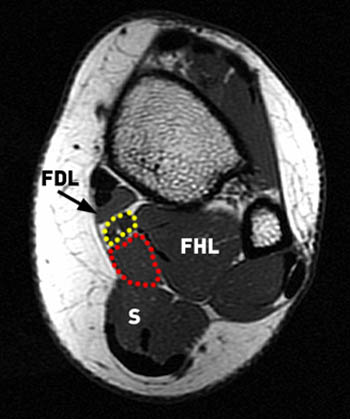
Abdm, abductor digiti minimi muscle;
Mri patterns of neuromuscular disease involvement thigh & other muscles 2. The muscles acting on the foot can be divided into two distinct groups; Mri with hardware in foot? Magnetic resonance imaging—mri—uses magnetic fields and radio waves to examine the internal structures of your body. Feet and ankles ankle muscle anatomy of foot muscles of foot muscles foot foot muscles anatomy muscle composite video showing multiple mri images including: The intrinsic foot muscles comprise four layers of small muscles that have both their origin and insertion attachments within the foot. A magnetic resonance imaging (mri) was performed on a normal subject; Learn about foot and ankle mri here. Head, neck, arm, foot, pelvis, etc. Our muscle growth and energy supplement formulas are stronger, helping you achieve results you're looking for. Indications for foot mri scan. Shop our pre workout and nitric oxide supplements. Case contributed by dr andrew dixon ◉.
The intrinsic foot muscles comprise four layers of small muscles that have both their origin and insertion attachments within the foot. Mri with hardware in foot? Case contributed by dr andrew dixon ◉. Learn more details about them at kenhub! This article reviews the use of magnetic resonance imaging (mri) in the evaluation of the foot, including a discussion of bone and cartilage abnormalities
Accessory Muscles of the Ankle - Radsource from radsource.us The intrinsic foot muscles comprise four layers of small muscles that have both their origin and insertion attachments within the foot. Related online courses on physioplus. This article reviews the use of magnetic resonance imaging (mri) in the evaluation of the foot, including a discussion of bone and cartilage abnormalities Learn about foot and ankle mri here. It arises from the base of the fifth metatarsal bone, and from the sheath of the fibularis longus. Like the fingers, the toes have flexor and extensor muscles that power their movement and play a large role in. Subscribe to foot & ankle problems. ► shoulder ► elbow ► wrist ► finger ► thumb.
Ankle mri (approach to msk mri series).
Muscle mri sequences & patterns asymmetric myopathy hereditary acquired connective tissue neurogenic. Indications for foot mri scan. Subscribe to foot & ankle problems. Learn about foot and ankle mri here. Intrinsic foot muscle weakness has been implicated in a range of foot deformities and disorders. Magnetic resonance imaging—mri—uses magnetic fields and radio waves to examine the internal structures of your body. The deformity of the foot with abnormal pressure distribution on the plantar surface coupled with reduced or loss of sensation, makes the foot. Head, neck, arm, foot, pelvis, etc. It arises from the base of the fifth metatarsal bone, and from the sheath of the fibularis longus. Ankle mri (approach to msk mri series). Shop our pre workout and nitric oxide supplements. Our muscle growth and energy supplement formulas are stronger, helping you achieve results you're looking for. Foot and ankle online course:
Share :
Post a Comment
for "Foot Muscles Mri / Foot, Ankle, and Calf | Musculoskeletal Key"


Post a Comment for "Foot Muscles Mri / Foot, Ankle, and Calf | Musculoskeletal Key"